Endovenous LASER Treatment for the elimination of varicose veins is quickly becoming the gold-standard in the treatment of varicose veins.
Endovenous LASER Treatment uses laser energy, which is simply a highly concentrated beam of light.
Medical lasers work by delivering this light energy to the targeted tissue with extreme precision, so as not to affect the surrounding tissue.
LASERs have proven their safety and effectiveness through years of use in all types of medical procedures, from eye surgery to dermatology.
In the hands of a skilled physician, lasers offer far less risk for complications than conventional surgery.
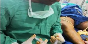
In endovenous laser treatment, a thin fiber is inserted into the damaged vein through a very small entry point in the skin.
A laser light is emitted through the fiber, as the fiber is pulled back through the vein, it delivers just the right amount of energy. The targeted tissue reacts with the light energy, causing the vein to close and seal shut.
The veins that are closed are superficial veins that handle less than five percent of the body’s blood flow. The blood is automatically routed to other, healthy veins.
The procedure is minimally invasive and requires no general anesthesia. Only local anesthetic is used to numb the area where the physician is working.
Patients are encouraged to walk immediately after the procedure and can resume normal activities the same day.
Endovenous LASER Treatment for the elimination of varicose veins is quickly becoming the gold-standard in the treatment of varicose veins.
Endovenous LASER Treatment uses laser energy, which is simply a highly concentrated beam of light.
Medical lasers work by delivering this light energy to the targeted tissue with extreme precision, so as not to affect the surrounding tissue.
LASERs have proven their safety and effectiveness through years of use in all types of medical procedures, from eye surgery to dermatology.
In the hands of a skilled physician, lasers offer far less risk for complications than conventional surgery.
In endovenous laser treatment, a thin fiber is inserted into the damaged vein through a very small entry point in the skin.
A laser light is emitted through the fiber, as the fiber is pulled back through the vein, it delivers just the right amount of energy. The targeted tissue reacts with the light energy, causing the vein to close and seal shut.
The veins that are closed are superficial veins that handle less than five percent of the body’s blood flow. The blood is automatically routed to other, healthy veins.
The procedure is minimally invasive and requires no general anesthesia. Only local anesthetic is used to numb the area where the physician is working.
Patients are encouraged to walk immediately after the procedure and can resume normal activities the same day.
A substantial change in medical practice has occurred in the last 10 years. Several new, minimally invasive techniques for treating have been described and come into general use. The main aim of these is to avoid surgical removal of varicose veins. The idea is to avoid the need for general anaesthetic, incisions in the legs and a hospital operating theatre leading to a much more rapid recovery from treatment. Ultrasound Guided Foam Sclerotherapy has become well established as a method of achieving long term cure of varicose veins.
A substantial change in medical practice has occurred in the last 10 years. Several new, minimally invasive techniques for treating have been described and come into general use. The main aim of these is to avoid surgical removal of varicose veins. The idea is to avoid the need for general anaesthetic, incisions in the legs and a hospital operating theatre leading to a much more rapid recovery from treatment. Ultrasound Guided Foam Sclerotherapy has become well established as a method of achieving long term cure of varicose veins.
At Scanroof Vein, palliative treatment is designed to relieve symptoms, and improve your quality of life. It can be used at any stage of an illness if there are troubling symptoms, such as pain or sickness. It can also be used to reduce or control the side effects of cancer treatments.
Some of the palliative treatment examples are as below:
Radiotherapy to reduce pain if cancer has spread to the bones.
Chemotherapy or targeted therapy to stop the cancer growing into other organs.
Surgery to reduce tumours causing pain or other symptoms.
Medicines to control symptoms and relieve discomfort.
What it is?
Hemorrhoids affect about 75% of the population by age 50. And, they are common in pregnancy. While hemorrhoids are not dangerous, they can bleed and become painful when they are in the skin around the anus.
A hemorrhoid is a cluster of swollen and inflamed veins in the lowest most part of the rectum.
The hemorrhoid may be internal, inside the lower rectum, and may protrude through the anus.
The only symptom may be bleeding after a bowel movement, or blood in the stool.
However, when the hemorrhoid protrudes it can collect mucus and microscopic amounts of stool that can cause external itching, pain and discomfort.
External hemorrhoids develop in the skin that surrounds the anus. Sometimes blood clots can occur in external hemorrhoids (Thrombosis).
This can cause bleeding, swelling and a hard lump around the anus. When the clot resolves extra skin is left behind and it can become irritated and itchy.
Rubbing and cleaning around the anus can make the symptoms worse.
Often the symptoms disappear after a few days, and some people are not aware they even have a hemorrhoid because they never have symptoms.
Diagnosis:
Because the symptoms of a hemorrhoid are similar to those of other anorectal problems like fissures, abscesses, warts and polyps, a physical exam and a digital rectal exam are employed to evaluate and diagnose hemorrhoids.
Any bleeding from the rectum requires a thorough exam to rule out other digestive diseases that can also cause bleeding. In people over the age of 40, a colonoscopy, sigmoidoscopy and barium enema x-rays may be used to rule out other causes, like colon cancer.
Medical treatment
Medical treatment for internal hemorrhoids include rubber band ligation, hemorroidectomy- cutting out the hemorrhoid, or staple hemorrhoidopexy.
What it is?
An anal fissure is a split or tear in the mucosa that lines the anus, that causes pain and bleeding with bowel movements.
They are most common in infants, and are less common in school aged children.
In adults, they may be caused by hard stool or diarrhea for an extended period. Additionally, a fissure may be due to decreased blood flow in older adults and high tension in the sphincter muscles.
And fissures are common in people with Crohn’s disease.
Diagnosis:
Anal fissures are diagnosed by a rectal exam and visualization of the area. But fissures are often confused with hemorrhoids.
Medical treatment
Treatment is aimed at softening stool and relaxing the anal sphincter to promote healing.
Most fissures heal on their own within 1-2 weeks. In infants, changing diapers often and cleaning the area can prevent and treat them.
Dietary changes to include more fiber and fluids, plus topical creams can soothe the area.
Chronic fissures are more difficult to treat than acute fissures, and can reoccur.
If the fissure does not heal with lifestyle improvements, a patient should seek medical help to assure there is no underlying disease.
Surgical options include Botox injection into the sphincter to relax it which will decrease pain and allow healing.
Botox treatment can heal 50-80% of patients. A procedure called a sphincterotomy is reported to be 90% effective.
What it is?
An abscess is an infected area near the anus that is filled with pus.
An anal fistula is commonly the result of a previous or current anal abscess. Small glands in the anus can get clogged and become infected, causing an abscess.
A fistula is a tunnel the forms under the skin that connects the infected glands to the abscess. But a fistula can develop without an abscess and connect the gland with the skin near the anus. Crohn’s disease, radiation, trauma and cancer can cause a fistula.
Diagnosis:
Most fistulas and abscesses are diagnosed by clinical findings. If there are deep abscesses or the fistula is not well delineated, imaging studies will help diagnose the situation.
Medical treatment
Treatment is surgical drainage of the abscess. Antibiotics may be prescribed in addition to surgical drainage. Surgery is necessary to cure a fistula.
Vacuum-assisted core biopsy is a safe and minimally invasive procedure in which a sample of breast tissue is removed for examination.
When breast imaging shows up very small abnormalities too small to be felt (i.e. anything unusual in the structure of the breast), Vacuum-assisted core biopsy is used to obtain samples of the breast tissue.
Vacuum-assisted core biopsy is an alternative to surgical biopsy. It allows the area of abnormality to be precisely located using imaging guidance so that only samples from the region of interest are removed.
At Scanroof Vein and Pain Clinic we have best in class facility available and expert doctor to perform this treatment. Get you appointment today to start with your treatment.
What it is?
Fine-needle aspiration (FNA) is a diagnostic procedure used to investigate lumps or masses. In this technique, a thin (23-25 gauge), hollow needle is inserted into the mass for sampling of cells that, after being stained, will be examined under a microscope (biopsy).
The sampling and biopsy considered together are called fine-needle aspiration biopsy (FNAB) or fine-needle aspiration cytology (FNAC).
When it is required?
This type of sampling is performed for one of two reasons:
A biopsy is performed on a lump or a tissue mass when its nature is in question.
For known tumors, this biopsy is performed to assess the effect of treatment or to obtain tissue for special studies.
An electrocardiogram (ECG) is a medical test that detects cardiac (heart) abnormalities by measuring the electrical activity generated by the heart as it contracts. … They may also recommend an ECG if a person is experiencing symptoms such as: chest pain. shortness of breath.
An electrocardiogram (ECG) is a medical test that detects cardiac (heart) abnormalities by measuring the electrical activity generated by the heart as it contracts. … They may also recommend an ECG if a person is experiencing symptoms such as: chest pain. shortness of breath.
An echocardiogram (echo) is a graphic outline of the heart’s movement. During an echo test, ultrasound (high-frequency sound waves) from a hand-held wand placed on your chest provides pictures of the heart’s valves and chambers and helps the sonographer evaluate the pumping action of the heart.
An echocardiogram (echo) is a graphic outline of the heart’s movement. During an echo test, ultrasound (high-frequency sound waves) from a hand-held wand placed on your chest provides pictures of the heart’s valves and chambers and helps the sonographer evaluate the pumping action of the heart.
Spirometry is a standard test doctors use to measure how well your lungs are functioning. Thetest works by measuring airflow into and out of your lungs.
Spirometry is a standard test doctors use to measure how well your lungs are functioning. Thetest works by measuring airflow into and out of your lungs.
OPG. An OPG is a panoramic or wide view x-ray of the lower face, which displays all the teeth of the upper and lower jaw on a single film. … An OPG may be requested for the planning of orthodontic treatment, for assessment of wisdom teeth or for a general overview of the teeth and the bone which supports the teeth.
OPG. An OPG is a panoramic or wide view x-ray of the lower face, which displays all the teeth of the upper and lower jaw on a single film. … An OPG may be requested for the planning of orthodontic treatment, for assessment of wisdom teeth or for a general overview of the teeth and the bone which supports the teeth.
A CT scan, also known as computed tomography scan, and formerly known as a computerized axial tomography scan or CAT scan, makes use of computer-processed combinations of many X-ray measurements taken
A CT scan, also known as computed tomography scan, and formerly known as a computerized axial tomography scan or CAT scan, makes use of computer-processed combinations of many X-ray measurements taken
Magnetic resonance imaging is a medical imaging technique used in radiology to form pictures of the anatomy and the physiological processes of the body in both health and disease. MRI scanners use strong magnetic fields, magnetic field gradients, and radio waves to generate images of the organs in the body.
Magnetic resonance imaging is a medical imaging technique used in radiology to form pictures of the anatomy and the physiological processes of the body in both health and disease. MRI scanners use strong magnetic fields, magnetic field gradients, and radio waves to generate images of the organs in the body.
Mammography is the process of using low-energy X-rays to examine the human breast for diagnosis and screening. The goal of mammography is the early detection of breast cancer, typically through detection of characteristic masses or microcalcifications.
Mammography is the process of using low-energy X-rays to examine the human breast for diagnosis and screening. The goal of mammography is the early detection of breast cancer, typically through detection of characteristic masses or microcalcifications.
Secondary Business TypeManufacturer / Exporters / Service Providers / Wholesale Suppliers
Opening Hours
SUN : Closed
MON : 9:30 AM - 6:30 PM
TUE : 9:30 AM - 6:30 PM
WED : 9:30 AM - 6:30 PM
THU : 9:30 AM - 6:30 PM
FRI : 9:30 AM - 6:30 PM
SAT : 9:30 AM - 6:30 PM
Causes of Varicose Veins Varicose veins Causes – The causes of varicose veins vary from person to person. The major reason for the cause of varicose veins could be due to weak or damaged valves. Some of the causes that can contribute to the risk of developing varicose veins are listed below. Heredity Obesity Lack of exercise Jobs requiring prolonged sitting or standing. High-fat