What is a labrum slap tear?
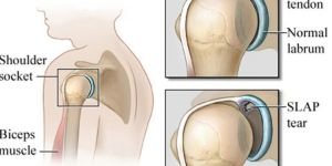
unless it causes you pain, you might never give your shoulder’s labrum a thought. This thick band of tissue surrounds your shoulder socket and keeps your shoulder joint stable. There are different kinds of labrum tears. A labrumslap tear happens in a specific area.
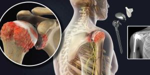
Your shoulder has three bones: the scapula (shoulder blade), humerus (upper arm bone), and clavicle (collarbone). They work together in a ball-and-socket joint where the arm connects to your trunk.
Your shoulder’s labrum isn’t a bone. It’s soft tissue that helps connect the socket part of the scapula (called the glenoid) with the head of the humerus. If the labrum tears, there’s not enough cushion between those bones. You also have a labrum in your hip sockets, which also acts like a connective cushion.
What 'slap' means
there are many different kinds of shoulder labrum tears. A labrum slap tear covers a specific area.
The upper, or superior, part of your labrum attaches to your biceps tendon. In a labrum slap tear, slap stands for superior labrum anterior and posterior. This means your labrum is torn at the top in both the front (anterior) and back (posterior) of where it attaches to the biceps tendon.
What is a labrum slap tear?
unless it causes you pain, you might never give your shoulder’s labrum a thought. This thick band of tissue surrounds your shoulder socket and keeps your shoulder joint stable. There are different kinds of labrum tears. A labrumslap tear happens in a specific area.
Your shoulder has three bones: the scapula (shoulder blade), humerus (upper arm bone), and clavicle (collarbone). They work together in a ball-and-socket joint where the arm connects to your trunk.
Your shoulder’s labrum isn’t a bone. It’s soft tissue that helps connect the socket part of the scapula (called the glenoid) with the head of the humerus. If the labrum tears, there’s not enough cushion between those bones. You also have a labrum in your hip sockets, which also acts like a connective cushion.
What 'slap' means
there are many different kinds of shoulder labrum tears. A labrum slap tear covers a specific area.
The upper, or superior, part of your labrum attaches to your biceps tendon. In a labrum slap tear, slap stands for superior labrum anterior and posterior. This means your labrum is torn at the top in both the front (anterior) and back (posterior) of where it attaches to the biceps tendon.
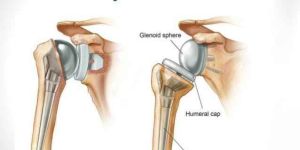
If your shoulder joint gets seriously damaged, you might need surgery to replace it. Before you have your procedure, you should know some things.
Dr. Nataraj H M is one of the Best Shoulder Replacement Doctor/Surgeon in Bangalore with over a Decade of Experience in Orthopedics and Shoulder Replacement.
Check out Below How Dr. Nataraj H M Describes about Shoulder Replacement Surgery’s expectations, Types of shoulder Replacement Surgeries, etc.
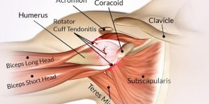
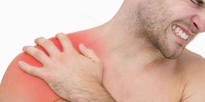
Shoulder impingement syndrome is a common cause of shoulder pain. It occurs when there is impingement of tendons or bursa in the shoulder from bones of the shoulder. An overhead activity of the shoulder, especially repeated activity, is a risk factor for shoulder impingement syndrome. Examples include painting, lifting, swimming, tennis, and other overhead sports. Other risk factors include bone and joint abnormalities.
With impingement syndrome, pain is persistent and affects everyday activities. Motions such as reaching up behind the back or reaching up overhead to put on a coat or blouse, for example, may cause pain.
Over time, impingement syndrome can lead to inflammation of the rotator cuff tendons (tendinitis) and bursa (bursitis). If not treated appropriately, the rotator cuff tendons can start to thin and tear.
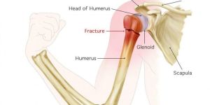
Shoulder fractures can result from a fall on the shoulder, a motor vehicle accident, contact sports, etc.
The shoulder is a complex joint connecting the arm to the body. The shoulder bones include the humerus (upper arm bone), the scapula (shoulder blade) and the clavicle (collarbone). The upper end of the humerus has a ball-like shape that connects with the socket of the scapula, called the glenoid. Disruption of any of the parts of the shoulder can create difficulty with its function.
In the past, instability was a lot different than it is now. In the 50s and the 60s, even in the 70s before we learned a lot about the shoulder, it was a traumatic anterior dislocation. “There were a few techniques and the measure of success was that you would have a stable shoulder. It wasn’t about the range of motion or playing sports; it was about the fact that your shoulder would never come out again.”
Since then, both the number of pathologies and operative choices has increased. “You can have anterior, posterior or multidirectional instability. We have new technologies, devices and instruments. Success is defined not only by stability but by mobility and actually returning to a level of function that our patients now demand from us.
Understanding the pathology of instability has evolved and has improved both surgical procedures. “I think we will come to a consensus that for anterior instability, the most important structure is the anterior inferior glenohumeral ligament, Dr Nataraj H M said. “The muscles play an extremely important role because they protect the capsule and the ligaments, but the repair has to be concentrated on reattaching the labrum if it is detached and tightening the capsule enough so that it is stable, yet you maintain a good range of motion.”
Shoulder Arthroscopy
Listed below is the step by step procedure of shoulder arthroscopy:
What is Shoulder Arthroscopy?
Why is Shoulder Arthroscopy Required?
Pre-operative Preparation
Day Before Surgery
Procedure Day
Methods/Techniques of Shoulder Arthroscopy
Post Procedure
Risks and Complications
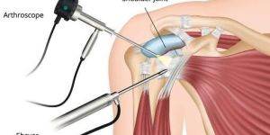
What is Shoulder Arthroscopy?
Shoulder arthroscopy is a minimally invasive procedure performed on the shoulder joint to repair its damaged portions. The surgery takes less than two hours to complete and is less painful than traditional open shoulder surgery. Also, the surgical procedure is minimally invasive, the recovery period is reduced. It is not a complex type of surgery and is a common treatment approach to cure severe shoulder pain.
Shoulder Arthroscopy
Listed below is the step by step procedure of shoulder arthroscopy:
What is Shoulder Arthroscopy?
Why is Shoulder Arthroscopy Required?
Pre-operative Preparation
Day Before Surgery
Procedure Day
Methods/Techniques of Shoulder Arthroscopy
Post Procedure
Risks and Complications
What is Shoulder Arthroscopy?
Shoulder arthroscopy is a minimally invasive procedure performed on the shoulder joint to repair its damaged portions. The surgery takes less than two hours to complete and is less painful than traditional open shoulder surgery. Also, the surgical procedure is minimally invasive, the recovery period is reduced. It is not a complex type of surgery and is a common treatment approach to cure severe shoulder pain.
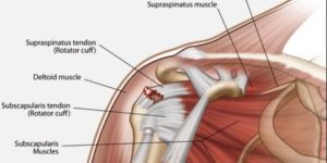
What Is Rotator Cuff Tear?
The rotator cuff is a combination of muscles and tendons that link your upper arm bone, the humerus, to your shoulder blades. The rotator cuff also holds your upper arm bone in place in your shoulder socket. The four muscles of the rotator cuff are the supraspinatus, the infraspinatus, the teres minor and the subscapularis. Each muscle is connected to the arm bone by a tendon. Rotator cuff repair is the surgery used to repair a tear in one of these tendons.
Symptoms of Rotator Cuff Injuries
People of all ages injure their rotator cuffs. Some are good candidates for surgery, while others will pursue different treatments.
You can injure your rotator cuff through wear and tear or poor movement patterns over time. Slouching and chronically pushing your head forward are two movement patterns that put your rotator cuffs at risk. As you get older, the rotator cuff can be irritated or pinched by calcium deposits in your shoulder area or bone spurs due to arthritis.
Repetitive stress is another culprit. Tennis players, swimmers, and baseball pitchers are at risk for repetitive stress injuries of the shoulder, as are carpenters and painters.
The knee can be divided into three compartments: patellofemoral, medial and lateral compartment. The patellofemoral compartment is the compartment in the front of the knee between the kneecap and thigh bone. The medial compartment is the area on the inside portion of the knee, and the lateral compartment is the area on the outside portion of the knee joint. Patellofemoral instability means that the patella (kneecap) moves out of its normal pattern of alignment. This malalignment can damage the underlying soft structures such as muscles and ligaments that hold the knee in place.
Causes
patellofemoral instability can be caused because of variations in the shape of the patella or its trochlear groove as the knee bends and straightens. Normally, the patella moves up and down within the trochlear groove when the knee is bent or straightened. Patellofemoral instability occurs when the patella moves either partially (subluxation) or completely (dislocation) out of the trochlear groove.
Knee resurfacing is an operative procedure that addresses localized cartilage (joint surface lining) defects in the knee joint and improves pain and function.
What is a femoral condyle cartilage defect?
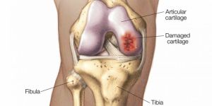
The knee joint consists of three bones; the femur; the tibia, and the patella or kneecap. The ends of the femur and tibia glide against each other as you move. Cartilage is the slippery shock absorbing material that covers the ends of bones to allow pain-free movement. A femoral condyle cartilage defect disrupts this surface which can lead to pain and loss of movement. If these defects are not treated they typically grow in size and depth until the pain is unmanageable.
Most of these defects are routinely treated by a technique called micro-fracture (drilling small holes in the bone) to promote some healing and eventually sealing off the defect with a layer of fibrocartilage.
Generally, in people aged between 35-65, sometimes these defects are quite large and the results with the microfracture technique are not very predictable or successful and in other people sometimes the micro-fracture treated areas fail and in both these situations using an implant to address the localized cartilage defect helps with pain and function.
Knee resurfacing is an operative procedure that addresses localized cartilage (joint surface lining) defects in the knee joint and improves pain and function.
What is a femoral condyle cartilage defect?
The knee joint consists of three bones; the femur; the tibia, and the patella or kneecap. The ends of the femur and tibia glide against each other as you move. Cartilage is the slippery shock absorbing material that covers the ends of bones to allow pain-free movement. A femoral condyle cartilage defect disrupts this surface which can lead to pain and loss of movement. If these defects are not treated they typically grow in size and depth until the pain is unmanageable.
Most of these defects are routinely treated by a technique called micro-fracture (drilling small holes in the bone) to promote some healing and eventually sealing off the defect with a layer of fibrocartilage.
Generally, in people aged between 35-65, sometimes these defects are quite large and the results with the microfracture technique are not very predictable or successful and in other people sometimes the micro-fracture treated areas fail and in both these situations using an implant to address the localized cartilage defect helps with pain and function.
Osteotomy literally means "cutting of the bone." In a knee osteotomy, either the tibia (shinbone) or femur (thighbone) is cut and then reshaped to relieve pressure on the knee joint.
Knee osteotomy is used when a patient has early-stage osteoarthritis that has damaged just one side of the knee joint. By shifting weight off of the damaged side of the joint, an osteotomy can relieve pain and significantly improve function in an arthritic knee.
Description
Osteoarthritis can develop when the bones of your knee and leg do not line up properly. This can put extra stress on either the inner (medial) or outer (lateral) side of your knee. Over time, this extra pressure can wear away the smooth articular cartilage that protects the bones, causing pain and stiffness in your knee.
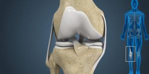
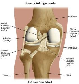
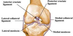
The knee joint is made to withstand the forces of walking, running and jumping as well as allowing motion. The knee is basically a hinge joint made of bone, ligament, and cartilage.
The anterior cruciate ligament or ACL is attached to the femur bone and tibia bone. The major function of the ACL is to provide stability to the knee during cutting and jumping activities. An ACL tear is a major problem for an athlete who desires pivoting, cutting and changing direction as part of their athletic lifestyle. These functions are required for sports such as skiing, soccer, football, basketball, volleyball and racquet sports. Once the ACL is torn, the knee becomes unstable and there is a risk of repeat injury and over time possibly advanced degeneration of the joint (arthritis).
The posterior cruciate ligament or PCL is one of the most important ligaments of the knee, giving it stability. The PCL achieves this role by preventing excessive twisting, straightening of the knee (hyperextension) and backward movement of the tibia on the femur.
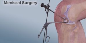
You have two c-shaped discs of cartilage (soft tissue) that connect your thighbone to your shinbone. These are called menisci. They’re like shock absorbers for your bones. They also help to keep your knee stable.
Athletes who play contact sports like football and hockey are prone to meniscus tears. But you can also get this injury when you kneel, squat, or lift something heavy. The risk of injury increases as you get older, when bones and tissues around the knee begin to wear down.
If you tear your meniscus, your leg might swell and feel stiff. You might feel pain when twisting your knee, or be unable to straighten your leg fully.
Ligaments are bands of tough, elastic connective tissue that surrounds a joint to give support and limit the joint's movement.
When ligaments are damaged, the knee joint may become unstable. Ligament damage often happens from a sports injury. A torn ligament severely limits knee movement. This results in the inability to pivot, turn, or twist the leg. Surgery is a choice to repair a torn ligament if another medical treatment is not effective.
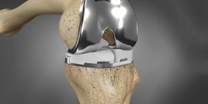
Total knee replacement is a complex surgical procedure wherein knee joint is replaced with prosthetic implant made up of artificial material or metals. Orthopedic surgeon takes precise measurements of the knee joint in order to determine the size of knee implant. Each component of knee is then replaced with metallic component and held in place with bone cement. The surgery takes around 2-3 hours to complete. Total knee replacement surgery is also called as total knee arthroplasty.
Why is total knee replacement surgery required?
total knee replacement surgery becomes necessary if:
arthritis or rare destructive knee joint diseases have severely damaged your knee
simple activities like walking, climbing stairs become very painful
sitting for long hours or lying down in one position also becomes painful
walking supports become of no use
medications and non-surgical treatments are useless to alleviate knee pain
knee joint has been so severely damaged due to accident or injury that it cannot be healed with non-surgical treatments
Knee Arthroscopy
Step by step procedure of knee arthroscopy:
What is Knee Arthroscopy?
Why is Knee Arthroscopy Required?
Pre-operative Preparation
Day Before Surgery
Procedure Day
Methods/Techniques of Knee Arthroscopy
Post Procedure
Risks and Complications
FAQs
What is Knee Arthroscopy?
It is a technique used for the treatment of knee joint problems. For diagnosis of the intra-articular knee disorders, such as synovial, meniscal, ligamentous and articular cartilage pathology, diagnostic arthroscopy is mandatory. The Mastery of diagnostic arthroscopy is important for orthopedic surgeons while treating knee disorders.
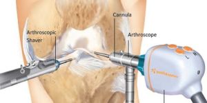
Knee arthroscopy is an important therapeutic procedure for treating knee disorders of various types. It is a surgical procedure which undertakes a small incision making and insertion of small camera called as arthroscope into the knees. Insertion of the arthroscope helps in observing the inside view of the joints on the screen of computer. It helps in identification of all knee problems. In some cases, problems knee can be corrected with the help of small instrument within the arthroscope.
Knee Arthroscopy
Step by step procedure of knee arthroscopy:
What is Knee Arthroscopy?
Why is Knee Arthroscopy Required?
Pre-operative Preparation
Day Before Surgery
Procedure Day
Methods/Techniques of Knee Arthroscopy
Post Procedure
Risks and Complications
FAQs
What is Knee Arthroscopy?
It is a technique used for the treatment of knee joint problems. For diagnosis of the intra-articular knee disorders, such as synovial, meniscal, ligamentous and articular cartilage pathology, diagnostic arthroscopy is mandatory. The Mastery of diagnostic arthroscopy is important for orthopedic surgeons while treating knee disorders.
Knee arthroscopy is an important therapeutic procedure for treating knee disorders of various types. It is a surgical procedure which undertakes a small incision making and insertion of small camera called as arthroscope into the knees. Insertion of the arthroscope helps in observing the inside view of the joints on the screen of computer. It helps in identification of all knee problems. In some cases, problems knee can be corrected with the help of small instrument within the arthroscope.
Frozen shoulder is also known as adhesive capsulitis. It usually involves stiffness and pain that develops gradually in your shoulder joint. Treatment for Frozen Shoulder involves stretching and sometimes injecting of corticosteroids and numbing medications into the joint capsule.
In some cases, surgery is used to loosen the joint capsule so that it can move freely. Signs and symptoms typically begin gradually, worsen over time and then resolve, usually within one or three years.
Symptoms of Frozen Shoulder
Frozen shoulder typically develops slowly, and in three stages. Each stage can last a number of months.
Freezing or Painful stage:Any movement of your shoulder causes pain, and your shoulder's range of motion starts to become limited.
Frozen stage:Pain may begin to diminish during this stage. However, your shoulder becomes stiffer, and using it becomes more difficult.
Thawing stage:The range of motion in your shoulder begins to improve.
For some people, the pain worsens at night, sometimes disturbing sleep.
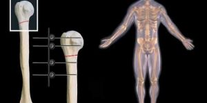
A broken bone must be carefully stabilized and supported until it is strong enough to handle the body's weight and movement. Until the last century, physicians relied on casts and splints to support and stabilize the bone from outside the body. The advent of sterile surgical procedures reduced the risk of infection, allowing doctors to internally set and stabilize fractured bones.
During a surgical procedure to set a fracture, the bone fragments are first repositioned (reduced) into their normal alignment. They are held together with special implants, such as plates, screws, nails, and wires.
Internal fixation allows shorter hospital stays, enables patients to return to function earlier, and reduces the incidence of nonunion (improper healing) and malunion (healing in improper position) of broken bones.
The implants used for internal fixation are made from stainless steel and titanium, which are durable and strong. If a joint is to be replaced, rather than fixed, these implants can also be made of cobalt and chrome. Implants are compatible with the body and rarely cause an allergic reaction.
Elbow arthroscopy
listed below is the step by step procedure of elbow arthroscopy:
what is elbow arthroscopy?
why is elbow arthroscopy required?
pre-operative preparation
day before surgery
procedure day
methods/techniques of elbow arthroscopy
post procedure
risks and complications
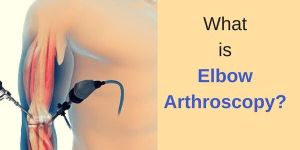
what is elbow arthroscopy?
elbow arthroscopy is an orthopaedic procedure that helps in the inspection, diagnosis and repairing of problems related to the joints of your elbow. Arthroscopy is a surgical procedure that involves making small incisions and inserting an arthroscope to look inside your joints. An arthroscope is a pencil-sized medical instrument having a source of light and a tiny camera attached to one of its ends, that helps to look inside your joints.
Elbow arthroscopy
listed below is the step by step procedure of elbow arthroscopy:
what is elbow arthroscopy?
why is elbow arthroscopy required?
pre-operative preparation
day before surgery
procedure day
methods/techniques of elbow arthroscopy
post procedure
risks and complications
what is elbow arthroscopy?
elbow arthroscopy is an orthopaedic procedure that helps in the inspection, diagnosis and repairing of problems related to the joints of your elbow. Arthroscopy is a surgical procedure that involves making small incisions and inserting an arthroscope to look inside your joints. An arthroscope is a pencil-sized medical instrument having a source of light and a tiny camera attached to one of its ends, that helps to look inside your joints.
This website focuses on articular cartilage repair treatment, which means the restoration of damaged hyaline cartilage in the joints.
Cartilage repair and regeneration is a treatment for joints that have damaged cartilage but are otherwise healthy.
The treatment is recommended for patients with cartilage damage or deterioration caused by:
Injury or trauma, including sports injuries
Repetitive use of the joint
Congenital abnormalities – abnormalities a person is born with – that affect a normal joint structure
Hormonal disorders that affect bone and joint development, such as osteochondritis dissecans (OCD)
There are several types of procedures for cartilage repair and regeneration that are designed to heal the cartilage by filling the cartilage defect (pothole) with repair tissue.
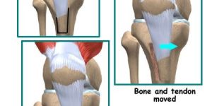
What is a biceps tenodesis?
A biceps tenodesis is a type of surgery used to treat a tear in the tendon that connects your biceps muscle to your shoulder. The tenodesis may be performed alone or as part of a larger procedure on the shoulder.
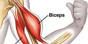
A tendon attaches muscle to bone. Your biceps tendons attach the biceps muscle of your upper arm to the elbow on one end and to the shoulder on the other. On the shoulder end, the biceps tendon divides into two strands, known as the long head and the short head.
The most common type of biceps tendon injury is in the long head biceps tendon (sometimes abbreviated as LHB).
What are the symptoms?
Biceps tendon tears may happen quickly from a traumatic injury or develop over time from repetitive motions of the shoulder.
Symptoms include:
a sudden, sharp pain in the upper arm, sometimes accompanied by a popping or snapping sound
cramping of the biceps during or after heavy use
pain or tenderness at the shoulder and elbow, or weakness in those areas
appearance of bruises from the middle of the bicep down toward the elbow
difficulty rotating the arm to a palm upward (or downward) position
a bulge in the upper arm, known as a “Popeye muscle”
What is a biceps tenodesis?
A biceps tenodesis is a type of surgery used to treat a tear in the tendon that connects your biceps muscle to your shoulder. The tenodesis may be performed alone or as part of a larger procedure on the shoulder.
A tendon attaches muscle to bone. Your biceps tendons attach the biceps muscle of your upper arm to the elbow on one end and to the shoulder on the other. On the shoulder end, the biceps tendon divides into two strands, known as the long head and the short head.
The most common type of biceps tendon injury is in the long head biceps tendon (sometimes abbreviated as LHB).
What are the symptoms?
Biceps tendon tears may happen quickly from a traumatic injury or develop over time from repetitive motions of the shoulder.
Symptoms include:
a sudden, sharp pain in the upper arm, sometimes accompanied by a popping or snapping sound
cramping of the biceps during or after heavy use
pain or tenderness at the shoulder and elbow, or weakness in those areas
appearance of bruises from the middle of the bicep down toward the elbow
difficulty rotating the arm to a palm upward (or downward) position
a bulge in the upper arm, known as a “Popeye muscle”
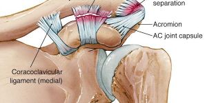
The main joint of the shoulder is a ball-and-socket joint formed by the glenoid socket and the humeral head. Most people think of this when they think of the shoulder joint. In fact, several joints make up the shoulder. The AC joint is a small joint directly on the top part of the shoulder and is the junction of the outer end of the clavicle (collarbone) and the acromion (upper part of the shoulder blade). A shoulder separation occurs when the acromion is forced away from the collarbone. Typically, it is forced up above the acromion resulting in a bony prominence on top of the shoulder. This usually happens as a result of a traumatic injury to the outer part of the shoulder. Common forms of this injury occurring in athletes include a hockey player going shoulder-first into the boards or a football player landing directly on the ground shoulder-first, often with another player on top, driving him into the ground. Non-athletic mechanisms can include falls onto the shoulder (often over the handlebars of a bike) or when trying to break a door down with one’s shoulder. The collarbone is held to the shoulder blade by two sets of ligaments, which often injured with a shoulder separation. If these ligaments are injured, the result is usually a higher grade or severity separation.
...more
Arthritis and AC Joint Dislocation Treatment Surgery
The main joint of the shoulder is a ball-and-socket joint formed by the glenoid socket and the humeral head. Most people think of this when they think of the shoulder joint. In fact, several joints make up the shoulder. The AC joint is a small joint directly on the top part of the shoulder and is the junction of the outer end of the clavicle (collarbone) and the acromion (upper part of the shoulder blade). A shoulder separation occurs when the acromion is forced away from the collarbone. Typically, it is forced up above the acromion resulting in a bony prominence on top of the shoulder. This usually happens as a result of a traumatic injury to the outer part of the shoulder. Common forms of this injury occurring in athletes include a hockey player going shoulder-first into the boards or a football player landing directly on the ground shoulder-first, often with another player on top, driving him into the ground. Non-athletic mechanisms can include falls onto the shoulder (often over the handlebars of a bike) or when trying to break a door down with one’s shoulder. The collarbone is held to the shoulder blade by two sets of ligaments, which often injured with a shoulder separation. If these ligaments are injured, the result is usually a higher grade or severity separation.
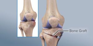
Reconstruction of the anterior cruciate ligament (ACL) is a surgery designed to restore knee stability and strength after the ligament has been torn. The remnants of the torn ligament must be removed and replaced with another ligament from your body or with tissue from a cadaver.
The knee is a hinge joint where the femur, or thighbone, meets the tibia, or shinbone.
in Bangalore Dr. Nataraj H M is one of the Best Orthopedic Doctor, Knee & shoulder Specialist in Bangalore with rich 13 years experience in Orthopedics. He has worked on clinical stem cell therapy for the treatment of arthritis of knee. Apart from regular orthopaedic work, he has been considered as one of the best Knee & Shoulder Pain Specialist in Bangalore India. He has Been Recognized for his Pioneering Work; to name a few- First Knee Surgeon in Southeast Asia to perform knee arthroscopic meniscal repair with Zimmer-Biomet meniscal repair device First Shoulder Surgeon in India to perform computer navigated (GPRS) total shoulder replacemen